

InciDENTALomas
What do you do when the image you take for one reason shows something unusual elsewhere on the image? As the number of diagnostic images taken increases, so to the likelihood of incidental findings.

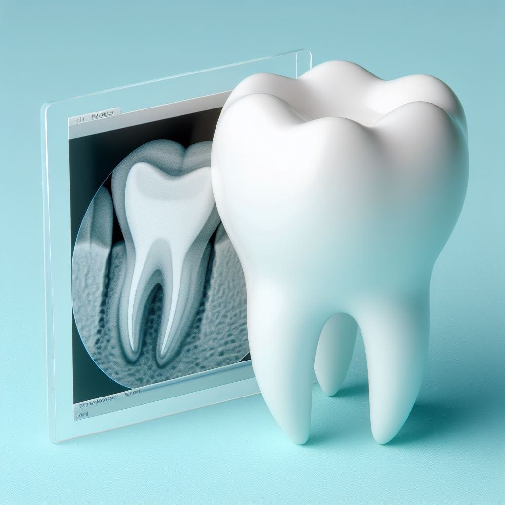
What do you do when the OPG or CBCT you take for a specific purpose shows something unusual somewhere else on the image? As the number of diagnostic images taken increases, so to the likelihood of incidental findings, which may pose dilemmas for both patients and practitioners.
A recent study in the Medical Journal of Australia highlighted concerns about incidental findings in up to 40% of patients undergoing diagnostic imaging scans for unrelated health conditions. This has doubled over the past 20 years and is likely due to technological advances in imaging technology and an increasing use in medical scans. For example, a brain CT scan used to consist of 24 slices, and that is now up to 240 slices. Abdominal and pelvis slices used to be 5mm thick, now they are down to 1mm, increasing the likelihood of incidental findings.
The problem is that whilst as few as 1% of these medical incidental findings may be deemed significant enough to warrant treatment, the findings invariably cause anxiety for patients, who may then undergo invasive procedures such as biopsies or radiation exposure for additional imaging to determine a diagnosis. This also leads to increased health care costs, and a strain on the health system for something that in most cases is of no medical consequence.
Of course, this is not confined to medicine. Improvements in dental imaging technology and a growing reliance on CBCT for a range of dental procedures is seeing a similar situation play out. There has been a significant increase in the number of CBCT and panoramic radiography machines in Australia over the past six years, and therefore a likely increase in the number of diagnostic images being taken. There were 706 CBCT machines and 3,059 panoramic radiation machines in 2020, an increase of 204% and 82% respectively since 2014.
A systematic review of the dental literature found a frequency of incidental findings of 24.6-94.3%, with common non-threatening findings including mucous retention cyst (55.1%), sinusitis (41.7%), soft tissue calcifications such as calcified stylohyloid ligament (26.7%), calcified pineal gland (19.2%), and tonsillolith (14.3%). Threatening incidental findings were much rarer – only about 1.4% of cases, although there were three papers reporting incidental carotid artery calcifications with a prevalence of 5.7–11.6%.
There is a need to consider the referral pathways for dental practitioners when incidental findings are not of a dental origin. What do you do, for example, when your imaging shows calcification of a coronary artery? It is critical that dental practitioners understand the implications of incidental findings and how they communicate that to their patients.
The increasing use of CBCT imaging has been mostly related to dental implant placement, but also for TMJ evaluation, orthodontic treatment, assessment of supernumerary teeth, endodontics and evaluating other pathology. It’s not clear whether the driver of this increased use is in the increased complexity of treatment provided (for example with dental implants), or whether it reflects dentists practising more defensively, with concerns about medico-legal risks driving a decision to use more sophisticated imaging ‘just in case.’ Or whether the increasing use is related to increased ownership of CBCT and panoramic radiation machines. Regardless of the reasons for the increasing use, dental practitioners need to be vigilant in their assessment of diagnostic images with an eye to these incidental findings.